
Haemoglobin SD disease results from a compound heterozygous state for haemoglobin S and haemoglobin D-Punjab. It is a severe sickling disorder with a moderate anaemia.
Haemoglobin S arises from a point mutation in the ß globin gene. Sickle cell mutations are more common in individuals of African descent. They are also found in Central and South America, the Middle East and parts of India.
Haemoglobin D-Punjab (also known as haemoglobin D-Los Angeles) also arises from a point mutation in the ß globin gene. It is one of the most common haemoglobin variants worldwide. Its prevalence in the Punjab region of India is estimated at 2%. It is also found in Italy, Turkey, Austria, Belgium, Brazil and Northwestern China.


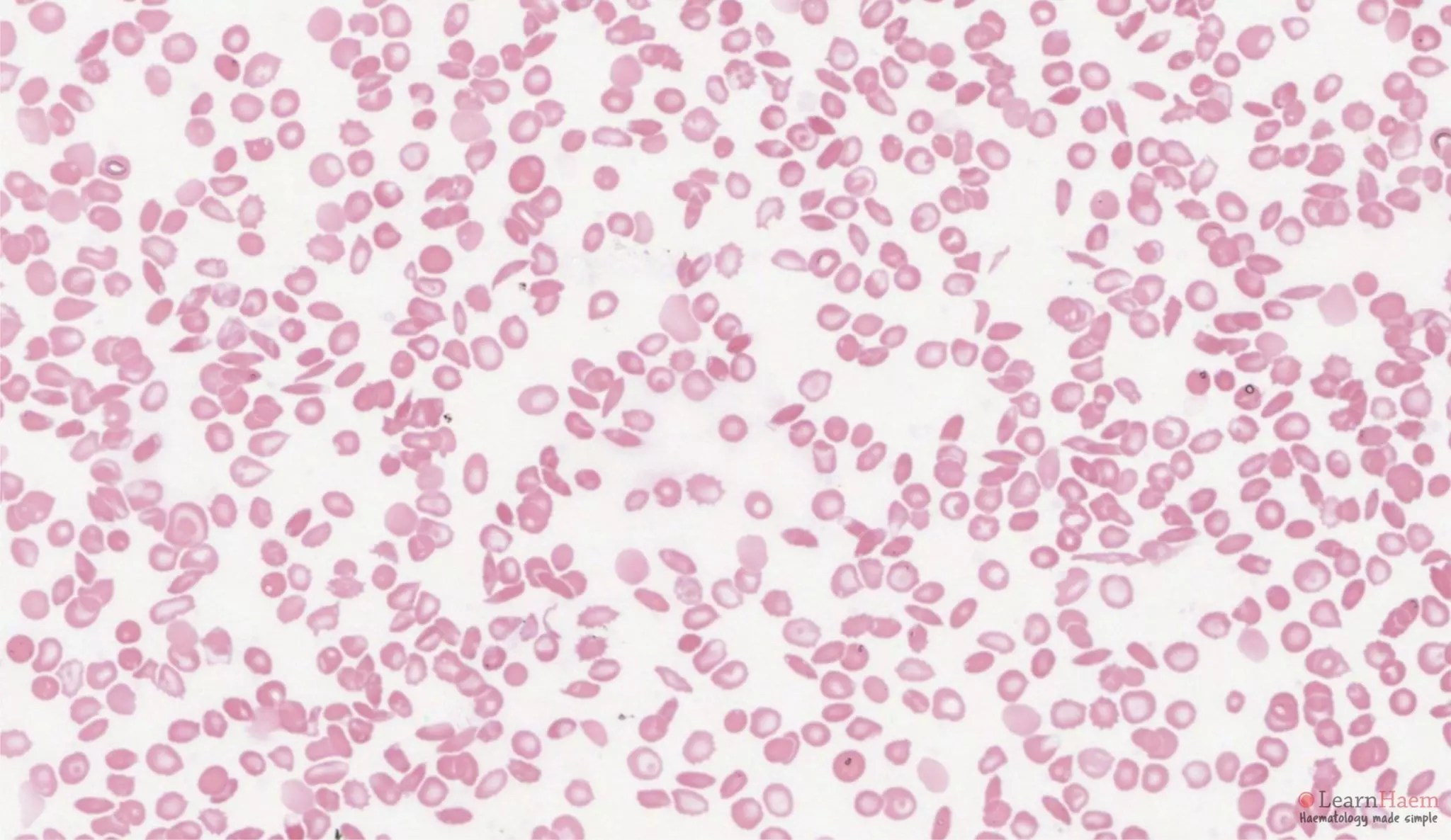
The blood film in a patient with haemoglobin SD disease resembles that of a patient with sickle cell disease. There is anisopoikilocytosis, target cells, sickle cells, boat cells and features of hyposplenism.
Haemoglobin Electrophoresis

Alkaline gel electrophoresis from a patient with haemoglobin SD disease. There are two variant haemoglobins, one with the mobility of haemoglobin S (red arrow) and the other with the mobility of haemoglobin F (blue arrow). The differentials for a band with the mobility of haemoglobin S are haemoglobins G, D and Lepore. The band at F represents HbF.

Acid gel electrophoresis from a patient with haemoglobin SD disease. There are two major variant haemoglobins. The first has the mobility of haemoglobin S (blue arrow), and represents haemoglobin S. The second has the mobility of haemoglobin A (red arrow), and represents haemoglobin D. Other differentials for a variant haemoglobin with the mobility of haemoglobin S on alkali gel but A on acid gel include haemoglobin G-Philadelphia (would not cause a sickling disorder) and haemoglobin Lepore (typically present in much smaller quantities).
HPLC
On HPLC, there will be no normal HbA. There will be two major variant haemoglobins, one with a retention time corresponding to haemoglobin S (~40%), and the other with a retention time corresponding to haemoglobin D (~50%). The remainder will be haemoglobin F (~2.5-5%) and haemoglobin A2 (2-3%).
Leave A Comment