
Sickle cell anaemia is an autosomal recessive disorder caused by a point mutation in the ß globin gene. It refers specifically to the sickling disorder which arises from the ßSßS genotype. Haemoglobin S is susceptible to deoxygenation, which triggers a conformational change that forms the basis for the pathogenic polymerisation of the variant haemoglobin.
The ßS gene is most often found in individuals of African descent, but can also be found in Mediterranean and Middle Eastern descent.


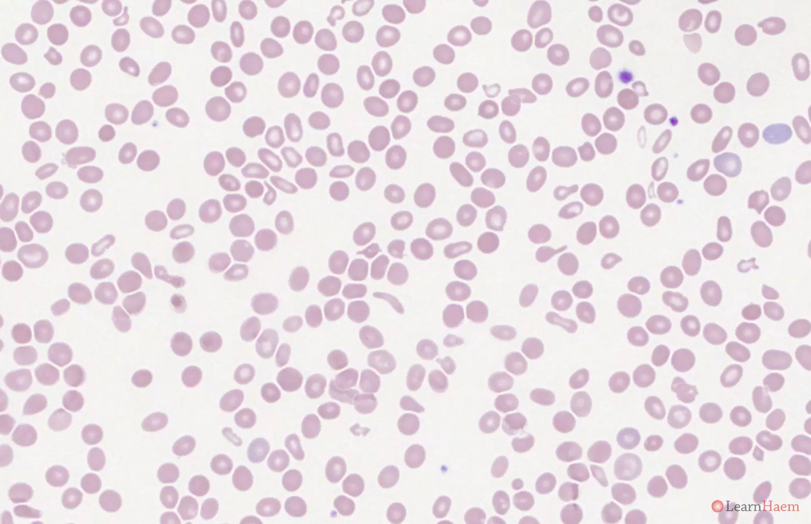
Blood film features:
- Sickle cells (may be very rare)
- Boat-shaped cells
- Polychromasia
- Anisocytosis
- Basophilic stippling
Other features to look for:
- Features of hyposplenism (although acanthocytes are rare in such patients)
- Malaria infection
- Dimorphic picture (recent transfusion)
- Features of megaloblastic anaemia (concurrent folate deficiency)
Differential diagnosis:
- Hb S / ß thalassaemia (more circulating nRBCs, basophilic stippling and other dyseythropoeitic features). The sickle test in both is positive.
- Haemoglobin SC disease (will have characteristic SC poikilocytes, anaemia is less severe, less dyserythropoeitic features). The sickle test in both is positive.
- ß thalassaemia intermedia / major (lacks sickle cells and boat-shaped cells). The sickle test in ß thalassaemia intermedia / major is negative.
Other investigations:
- Sickle test: positive in Hb S / ß thalassaemia
- Haemoglobin electrophoresis
- HPLC
Haemoglobin electrophoresis:

Alkaline gel electrophoresis from a patient with sickle cell anaemia. The main haemoglobin has the mobility of haemoglobin S. There are other haemoglobins with the mobility of haemoglobin F and haemoglobin C. The band with the mobility of haemoglobin C is most likely haemoglobin A2.

Acid gel electrophoresis from a patient with sickle cell anaemia. The main haemoglobin has the mobility of haemoglobin S. This confirms that the dominant band seen on the alkaline gel is haemoglobin S (Hb G, D and Lepore all have the mobility of Hb A on acid gel). There are other haemoglobins with the mobility of haemoglobin F and haemoglobin A. The band with the mobility of haemoglobin A is most likely haemoglobin A2.
HPLC:
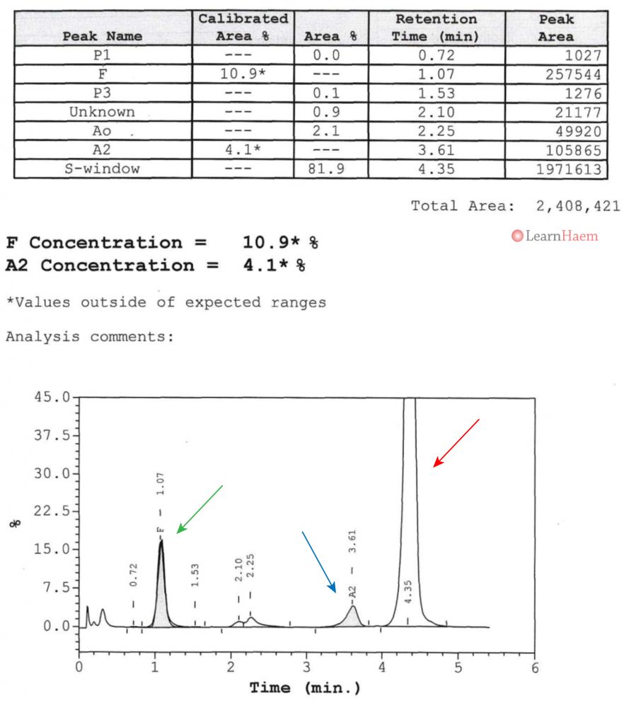
HPLC from a patient with sickle cell anaemia. There is no normal haemoglobin A. The major haemoglobin is haemoglobin S (red arrow). The haemoglobin F (green arrow) fraction is increased (10.9%, upper limit of normal usually ~1%), which helps to compensate for the severity of the disease. The low concentration of the variant haemoglobin in the A2 window (blue arrow) confirms that it is haemoglobin A2 (haemoglobin E can also occur in this window, but would be expected to have a higher fraction, with a lower fraction of Hb S).
Other resources:
- PHE Family Origin Questionnaire
- PHE Sickle Cell and Thalassaemia Screening Programme (2019)
- BCSH Guideline: Significant Haemoglobinopathies: Guidelines for Screening and Diagnosis (2010)
- BCSH Guideline: Management of Acute Chest Syndrome in SCD (2015)
- BCSH Guideline: Red Cell Transfusion in SCD Part I (2016)
- BCSH Guideline: Red Cell Transfusion in SCD Part II (2016)
- BCSH Guideline: Use of hydroxycarbamide in children and adults with SCD (2018)
- BCSH Guideline: Red blood cell specifications for patients with hemoglobinopathies: a systematic review and guideline (2020)
- RCOG Guideline: Management of SCD in Pregnancy (2011)
Leave A Comment