

Clinical features
- Males more commonly affected (2.5 : 1)
- Inflammatory back pain
- Typically starts in third or fourth decade of life
- Insidious onset
- Worse in the morning or after periods of immobility
- Better with exercise
- May have difficulty sleeping
- Not relieved by resting
- May have buttock pain (sacroiliac involvement)
- Can have symptoms anywhere along the spine
- Relieved by NSAIDs
- Family history of spondyloarthropathy
- Other manifestations
- Peripheral arthritis
- Enthesitis
- Dactylitis
- Anterior uveitis (acute, unilateral painful red eye with photophobia, blurring of vision)
- Aortic regurgitation (sclerosing inflammation resulting in decreased elasticity of the aortic root)
- Extra-Athoracic restrictive lung disease (diminished chest wall and spinal mobility)
- Apical pulmonary fibrosis
- Spinal involvement
- Modified Schober test (anterior lumbar spinal flexion)
- Midpoint of the line joining the two posterior superior iliac spines
- Note the position 10cm above and 5cm below this point with the patient standing
- Ask the patient to bend forward as much as possible, while keeping legs straight
- Distance between the two points should increase by ≥ 5cm
- Lateral cervical and lumbar spinal flexion
- Occiput to wall distance (normal people should be able to touch occiput to wall when standing)
- Cervical spine and thoracic spine rotation
- Modified Schober test (anterior lumbar spinal flexion)
- Complications
- Spinal cord injury secondary to pathological fractures
- Atlanto-axial subluxation
- Cauda equina syndrome
- Commonly co-exists with
- Psoriasis
- Inflammatory bowel disease
Investigations
- Full blood count (anaemia of chronic disease)
- Renal function, liver function (prior to starting NSAIDs or definitive therapy)
- HLA-B27 (positive in 90% of patients with AS, but not specific)
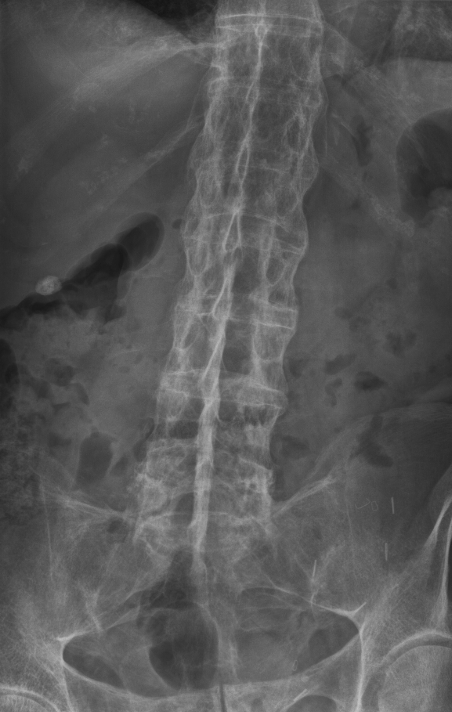
- Plan radiograph of the pelvis and spine
- Scaroilitis
- Erosions, osteitis at bony prominences
- Presence of syndesmophytes (bony growths within ligaments) – bamboo spine
- Magnetic resonance imaging of sacroiliac joints to look for active inflammation
- Trans-thoracic echocardiogram to assess aortic valve if history and examination suggestive
- High-resolution computed tomography scan of the lungs to look for apical fibrosis
- Pulmonary function testing to look for restrictive lung disease
Management
- NSAIDs (first-line treatment): continuous use can slow radiographic progression
- Conventional DMARDs have no role in the treatment of AS (they are not effective)
- TNF-α inhibitors
- Indicated in patients who have inadequate response to at least two NSAIDS used for ≥4 weeks each
- Choices: etanercept, adaliumumab, golimumab, infliximab
- > 60% response rate
- May switch to a different TNF-α inhibitor if response to the first decreases
- Reduces radiographic progression of AS
- Rituximab (anti-CD20)
- May have some efficacy in TNF-α naïve patients
- Currently role in AS is unclear
- Newer targets
- Anti-IL12/23: ustekinumab
- Anti-IL17: secukinumab
CVS: aortitis, conduction disturbances and cardiomyopathy