


Clinical features
- Non-proliferative diabetic retinopathy (NPDR)
- No neovascularization
- Mild
- ≥ 1 microaneurysms
- Moderate
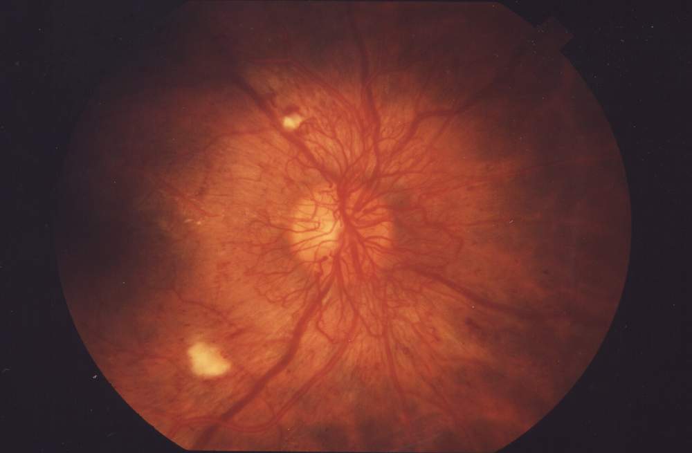
- Soft exudates (cotton wool spots) – nerve fibre layer infarction
- Hard exudates – lipid deposition
- Haemorrhages (dot or blot)
- Venous beading
- Severe
- Venous beading in at least two quadrants
- Proliferative diabetic retinopathy (PDR)
- Neovascularization from the disc or retinal vessels
- May arise in the absence of NPDR
- Early
- New vessels
- High-risk
- Macula involved
- Complications
- Vitreous haemorrhage
- Retinal detachment
- Macula oedema
- May occur at any stage of DR
- Retinal thickening
- Seen with fluorescein angiography
- DR is usually asymptomatic until very late
- Usually not reversible by symptomatic stage
- Require regular screening
Exacerbating factors
- Pregnancy
- Poor glycaemic control
- Hypertension
- Diabetic nephropathy
- Thigh glycaemic control in patients with previously poor glycaemic control
Ocular manifestations of diabetes
- Central retinal artery occlusion
- Central retinal vein occlusion
- Mononeuritis multiplex
- Vitreous haemorrhage
- Retinal detachment
- Cataracts
Indications for urgent referral (within 2 weeks)
- Proliferative diabetic retinopathy
- Macular involvement
- CRAO / CRVO
- Retinal detachment
- Vitreous haemorrhage
Leave A Comment