
History
- Sudden onset
- Painless loss of vision or blurred vision
Physical examination
- Relative afferent pupillary defect
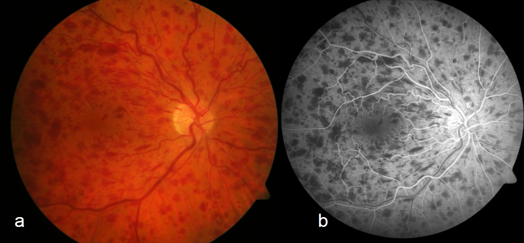
- Fundoscopy
- Scattered, diffuse retinal haemorrhage
- “Blood and thunder” fundus
- Dilated, tortuous veins
- Optic disc oedema
- Cotton wool spots – retinal ischaemia
- Arterioles may be attenuated
Risk factors
- Age
- Hypertension
- Diabetes
- Smoking
- Obesity
- Glaucoma – affects retinal vein outflow
- Hypercoagulable states
- Antiphospholipid syndrome
- Nephrotic syndrome
- Factor V Leiden
- Protein C deficiency
- Protein S deficiency
- Hyperhomocysteinaemia
- Underlying malignancy
Complications
- Ocular neovascularization
- Vitreous haemorrhage
- Traction retinal detachment
- Neovascular glaucoma
Management
- Multidisciplinary team approach
- Refer to ophthalmology
- Control cardiovascular risk factors
- Consider intra-viteal anti-vascular endothelial growth factor (VEGF) for neovascularization with macular oedema
- Laser phototherapy for neovascularization
- Consider intravitreal glucocorticoids (second-line)
Leave A Comment