
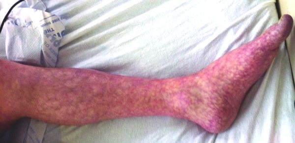
Erythema Nodosum
Characteristics

- Painful, symmetrical red nodules
- Usually 1 – 10 cm in size
- Most often on anterior legs
- Involutes in weeks, giving a bruise-like appearance
- New lesions may occur over up to eight weeks
- Does not ulcerate, tends to heal completely
- May have prodromal illness
- Malaise
- Fever
- Arthralgia
- Histology: septal panniculitis
Differential diagnosis
- Infective
- Streptococcal pharyngitis
- Tuberculosis
- Hepatitis B and C
- Epstein-Barr virus
- Human immunodeficiency virus
- Whipple’s disease
- Gastroenteritis secondary to bacterial infection
- Histoplasmosis
- Syphilis
- Inflammatory
- Sarcoidosis (Löfgren’s syndrome – acute sarcoidosis with EN, bilateral hilar lymph nodes, arthritis)
- Inflammatory bowel disease
- Behcet’s syndrome
- Malignancy
- Hodgkin’s lymphoma
- Drugs
- Antibiotics
- Sulphonamides
- Amoxicillin
- Oral contraceptive pill
- Montelukast
- Antibiotics
- Neoplastic
- Lymphoma
- Pregnancy
- Idiopathic (up to 55%)
Investigations
- Confirm diagnosis: biopsy of skin lesion
- Full blood count to look for evidence of infection, differential to screen for lymphoproliferative disease
- Liver function tests (hepatitis B and C)
- Human immunodeficiency virus test
- Chest radiograph
- Bilateral hilar lymphadenopathy (sarcoidosis)
- Apical consolidation (tuberculosis)
- Sputum for acid-fast bacilli stain and culture, molecular detection of mycobacterium tuberculosis
- Consider colonoscopy to evaluate for ulcerative colitis
- Consider pharyngeal swab for Group A Streptococci, anti-streptolysin-O titre
- Consider stool culture for gastrointestinal organisms
Management
- Multidisciplinary team approach
- Patient education: tends to be self-limiting
- Consider referral to Dermatology
- Analgesia: paracetamol, non-steroidal anti-inflammatory drugs
- Treat underlying cause
- If severely symptomatic, consider oral potassium iodide 400 – 900mg / day (most likely to be effective if started early)
- Monitor thyroid function closely
- May also consider prednisolone 1mg/kg/day if very symptomatic
Credit: James Heilman, MD
Erythema Multiforme

Characteristics
- Acute
- Immune-mediated
- Lesions may have mucosal involvement
- Acrally distributed papules surrounded by erythema
- Distinct, target lesions
- Concentric colour changes
- Usually self-limiting, but may be recurrent
- Recent consensus classification:
- Distinct from Stevens-Johnson syndrome
Differential diagnosis
- Infections
- Herpes simplex virus (usually type 1)
- Mycoplasma pneumoniae
- Hepatitis C
- Epstein-Barr virus
- Drugs
- Sulphonamides (Bactrim)
- Sulphonylureas
- Anti-epileptics
- Antibiotics
- Non-steroidal anti-inflammatory drugs
- Allopurinol
- Inflammatory
- Inflammatory bowel disease
- Graft versus host disease
- Polyarteritis nodosa
- Sarcoidosis
- Malignancy (leukaemia and lymphoma)
Investigation
- HSV PCR from suspicious skin lesions
- Full blood count
- Liver function tests (hepatitis C)
- Chest radiograph (Mycoplasma, sarcoidosis)
- Consider serological test for Mycoplasma
- Consider colonoscopy (inflammatory bowel disease)
- Consider skin biopsy
- Consider age-appropriate malignancy screening
Management
- Patient education
- Stop offending drug, if any
- Treat underlying disease
- Symptomatic treatment: antihistamines for pruritus
- Consider topical corticosteroids if symptomatic
- Consider antiviral prophylaxis or dapsone for recurrent EM
Livedo Reticularis
Characteristics
- Increased visibility of venous plexuses
- May be caused by
- Deoxygenation
- Decreased flow
- Increased viscosity
- Increased venous resistance
- Venous thrombosis
- Venodilation
- Local hypoxia
- Dysautonomia
- Deoxygenation
- Mottled, reticulated vascular pattern
- Lace-like purplish discolouration of skin
Differential diagnosis
- Inflammatory
- Anti-phospholipid syndrome
- Systemic lupus erythematosus
- Sneddon’s syndrome (LR + cerebrovascular lesions)
- Hyperviscosity
- Cryoglobulinaemia (may be secondary to hepatitis C)
- Cold agglutinin disease (may be secondary to Mycoplasma infection)
- Polycythaemia vera
- Essential thrombocytosis
- Acute leukaemia
- Chronic lymphocytic leukaemia
- Waldenström’s macroglobulinaemia
- Embolic disease
- Cholesterol embolisation syndrome
- Septic emboli
- Hypercoagulable states
- Anti-thombin III deficiency (may be secondary to nephrotic syndrome)
- Protein C deficiency
- Protein S deficiency
- Drugs
- Amantadine
- Minocycline
- Gemcitabine
- Warfarin (skin necrosis)
- Calciphylaxis (may be secondary to chronic kidney disease)
- Congenital
- Cutis marmorata telangiectasia congenita
- Ehlers-Danlos syndrome
Relevant points in the history
- Anti-phospholipid syndrome
- Have you ever had blood clots in your legs or lungs?
- Have you ever had a stroke?
- Have you ever had a heart attack?
- Have you ever had any miscarriages?
- Systemic lupus erythematosus
- Have you lost any hair?
- Do you have any mouth ulcers?
- Do you get any joint pains?
- Have you ever had a rash on your face?
- Hepatitis C
- Have you ever had a blood transfusion?
- Have you ever injected drugs through a vein?
- Have you ever noticed episodes where your skin or eyes have appeared yellow?
- Have you noticed a rash on your chest or face?
Investigations
- Full blood count (anaemia of chronic disease, myeloproliferative disease)
- Auto-antibodies
- Anti-phospholipid syndrome
- Anti-cardiolipin (IgG and IgM)
- Anti-β2 glycoprotein (IgG and IgM)
- Lupus anticoagulant
- Systemic lupus erythematosus
- Anti-nuclear
- Anti-double stranded DNA
- Renal function
- Calcium (calciphylaxis)
- May consider skin biopsy if diagnosis uncertain
- Anti-phospholipid syndrome
Management
- Consider low-dose aspirin if patient asymptomatic
- If previous thrombosis: consider anticoagulation (lifelong)
- Treat the underlying cause
Easy Bruising
Differential diagnosis
- Cushing’s syndrome
- Henoch-Schonlein purpura
- Connective tissue disease
- Marfan’s syndrome
- Ehlers-Danlos syndrome
- Platelet abnormalities
- Immune thrombocytopaenic purpura
- von Willebrand’s disease
- Coagulopathy
- Chronic liver disease
- Vitamin K deficiency
- Congenital haemophilia
- Acquired haemophilia
- Acute promyeloid leukaemia
Hypopigmentation
Differential diagnosis
- Vitiligo
- Tinea vesicolour
- Ash leaf spots (tuberous sclerosis)
- Leprosy
- Sarcoidosis
- Cutaneous T cell lymphoma
- Depigmentation associated with underlying melanoma
Characteristics of vitiligo
- Acquired depigmentation of the skin
- Autoimmune process directed at melanocytes
- Onset usually in second and third decade
- Usual distribution is acral
- May display Koebner phenomenon
- Usually slowly progressive
- 10 – 20% may experience spontaneous re-pigmentation
Associated autoimmune disorders
- Hyperthyroidism (Graves’)
- Hypothyroidism (Hashimoto’s)
- Addison’s disease
- Pernicious anaemia
- Systemic lupus erythematosus
- Psoriasis
- Rheumatoid arthritis
- Inflammatory bowel disease
- Autoimmune polyglandular syndrome type II
- Autoimmune thyroid disorders
- Type I diabetes mellitus
- Hypopituitarism
- Primary adrenal insufficiency
Examination
- Vitals, including postural blood pressure
- Thyroid status
- Tremor
- Pulse
- Coarse facies
- Coarse hair
- Hoarse voice
- Goitre
- Proximal myopathy
- Pretibial myxoedema
- Examination of skin
- Psoriasis – especially nails, elbows, scalp, retro-auricular
- Melanoma
- Inflammatory disorders
- GALS screen
- Rheumatoid nodules
- Mouth ulcers
- Ideally: examine vitiligo under Wood’s lamp to differentiate depigmentation from lightly-pigmented skin
Investigations
- Consider fasting plasma glucose
- Consider thyroid function tests
- Consider anterior pituitary hormone screen
- Consider short synACTHen tests
Management
- Sunscreen to minimize tanning and contrast between normal skin and depigmented skin
- Makeup (e.g. Dermablend) to camouflage depigmented areas
- Topical corticosteroids (moderate quality evidence for short-term benefit)
- Ultraviolet therapy (moderate evidence)
Leave A Comment