

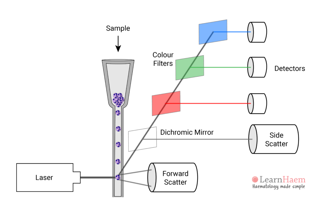
Modern flow cytometers can process thousands of cells per second. The basic premise of all flow cytometers is that when beam of light is directed at an object, the light will scatter; this scatter depends on the granularity and opacity of the object. In flow cytometry, laser beams provide the light source, while cells in single file are presented as obstacles; varying signals are generated depending on the size, internal complexity and granularity of the cell.
There are three main components to most flow cytometers:
- Fluidics system: cells presentation to lasers.
- Optical system: collecting information from light scatter of cells. The main types of photodetectors used are photodiodes and photomultiplier tubes, which are more efficient and better for collecting weaker signals.
- Electronics system: conversion of optical to digital signals for display and analysis/interpretation. Light signals are converted into voltage pulses, the magnitude of which is proportional to the number of photons detected. These are most often displayed as dot plots, with each dot representing a single cell.
Leave A Comment